Visiting UBC’s Materials Engineering Department
Few disciplines pay attention to materials as closely as Materials Science and Engineering. The Animate Materials Workshop summer 2026 research team visited the University of British Columbia’s Department of Materials Engineering to deepen our understanding of materials through engagement with scientific research and visualization methods. Our visit included a brief tour of the Frank Forward Building, which houses the Electron Microscopy Laboratory led by Dr. Ben Britton.
-
Exterior of the Frank Forward Building at the University of British Columbia.
-
Closer look at the building’s brickwork mural designed by sculptor George Norris.
-
Alchemical symbols displayed inside the building.
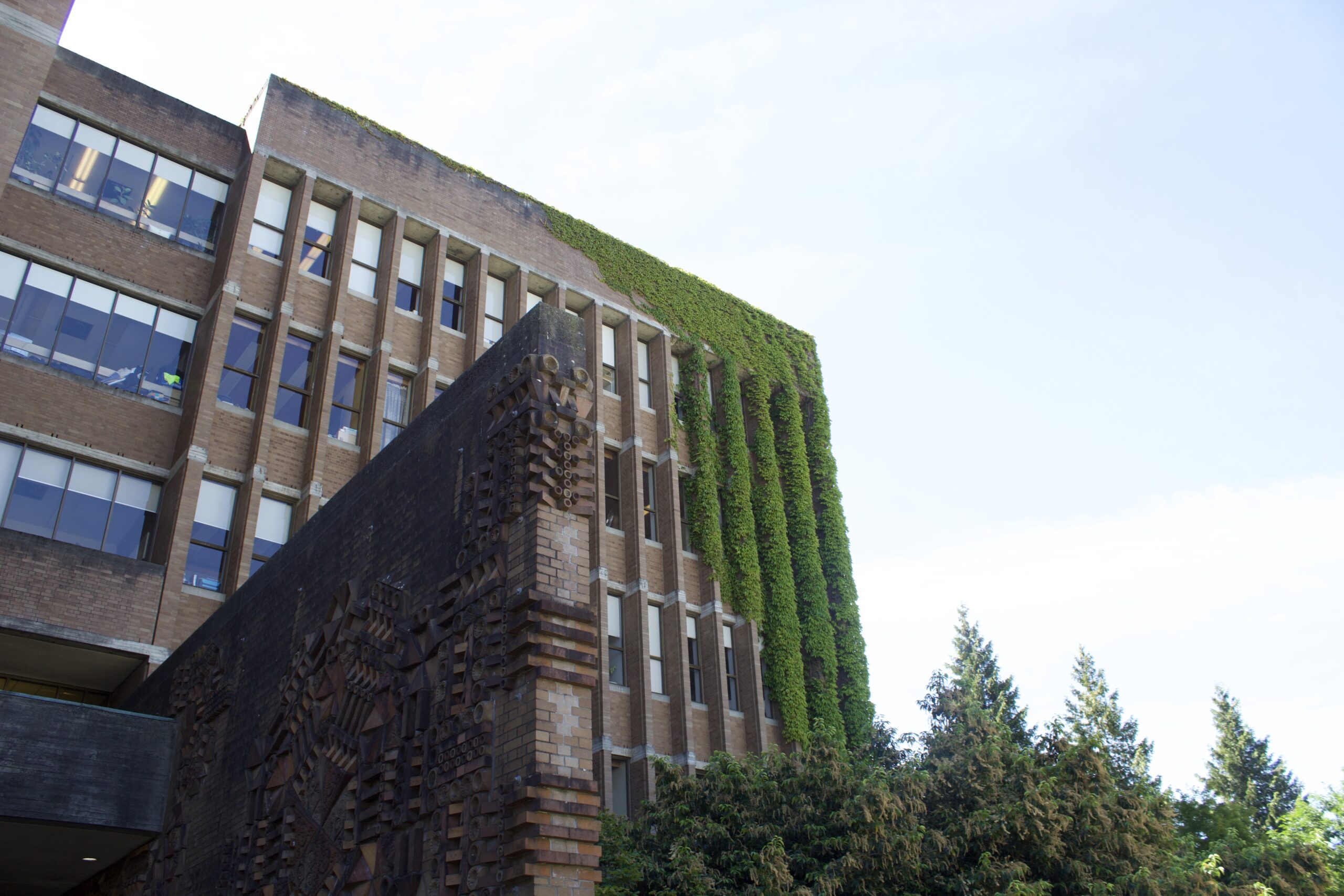
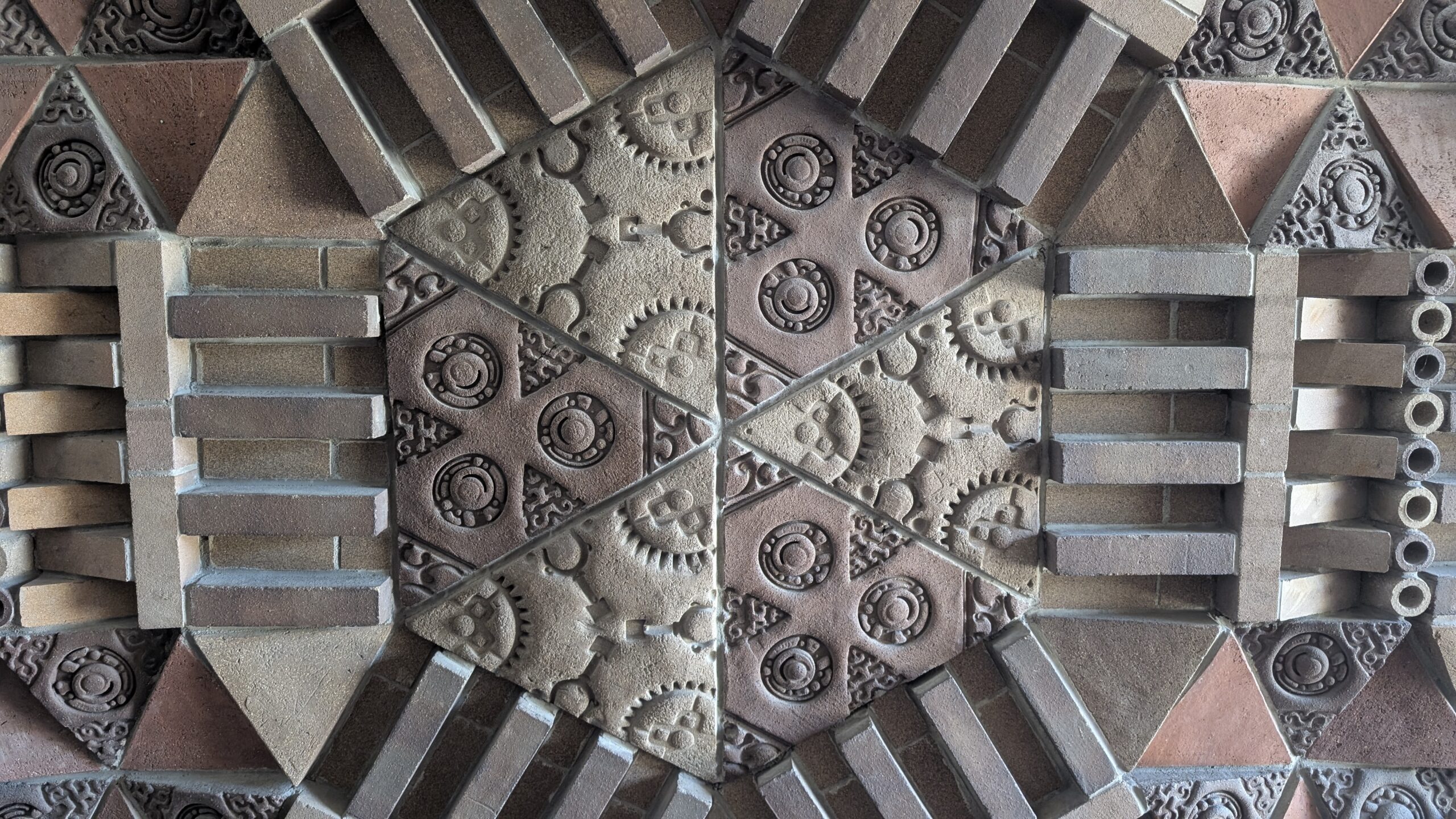
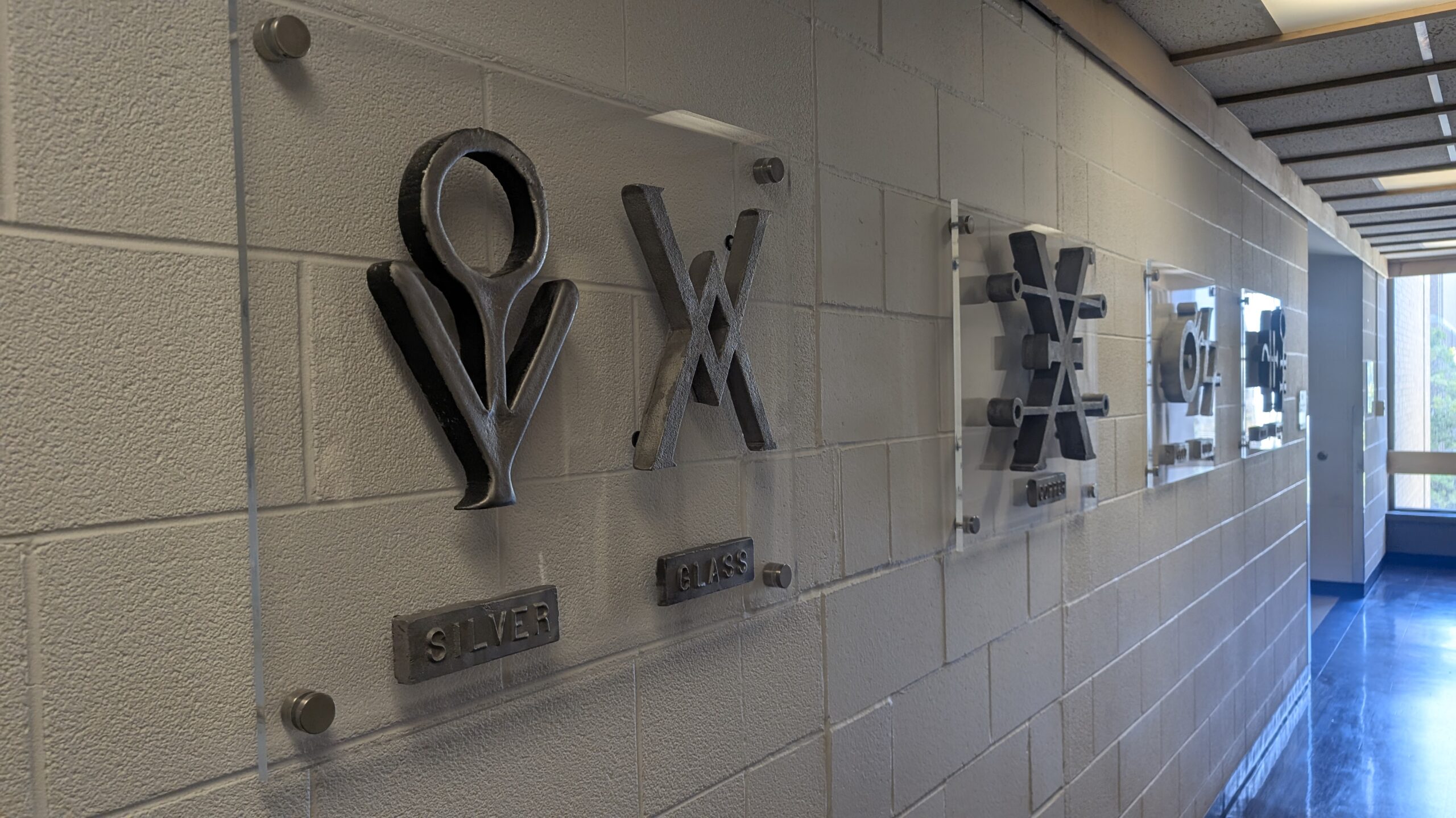
The Department of Metallurgy (now the Department of Materials Engineering) was among the first programs established in 1915, following UBC’s founding under the 1908 University Act. During the late 1890s and early 1900s, mining booms in the Kootenays and Boundary districts generated an urgent demand for skilled metallurgists and mining engineers. Emphasizing Applied Science as central to UBC’s mission helped persuade lawmakers that the university would directly increase provincial industrial output. The Frank Forward Building was an important emblem of the university’s heritage, commemorated in an impressive brick-laid artwork designed by BC sculptor George Norris. This abstract mosaic draws inspiration from the crystalline atomic structure of metallic compounds. Norris made the bricks himself, pressing metallic components onto their surfaces before firing them to create their intricate designs. We were curious to see the artwork displayed throughout the building, including rubbings of medieval architectural reliefs and a wall dedicated to alchemical symbols, the early history of modern materials science.
-
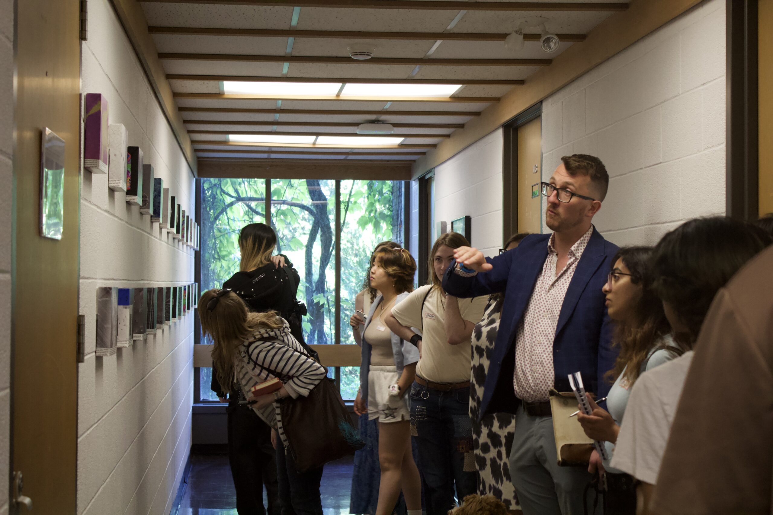
Dr. Ben Britton led the tour for the AMW participants.
-
Dr. Ben Britton led the tour for the AMW participants.
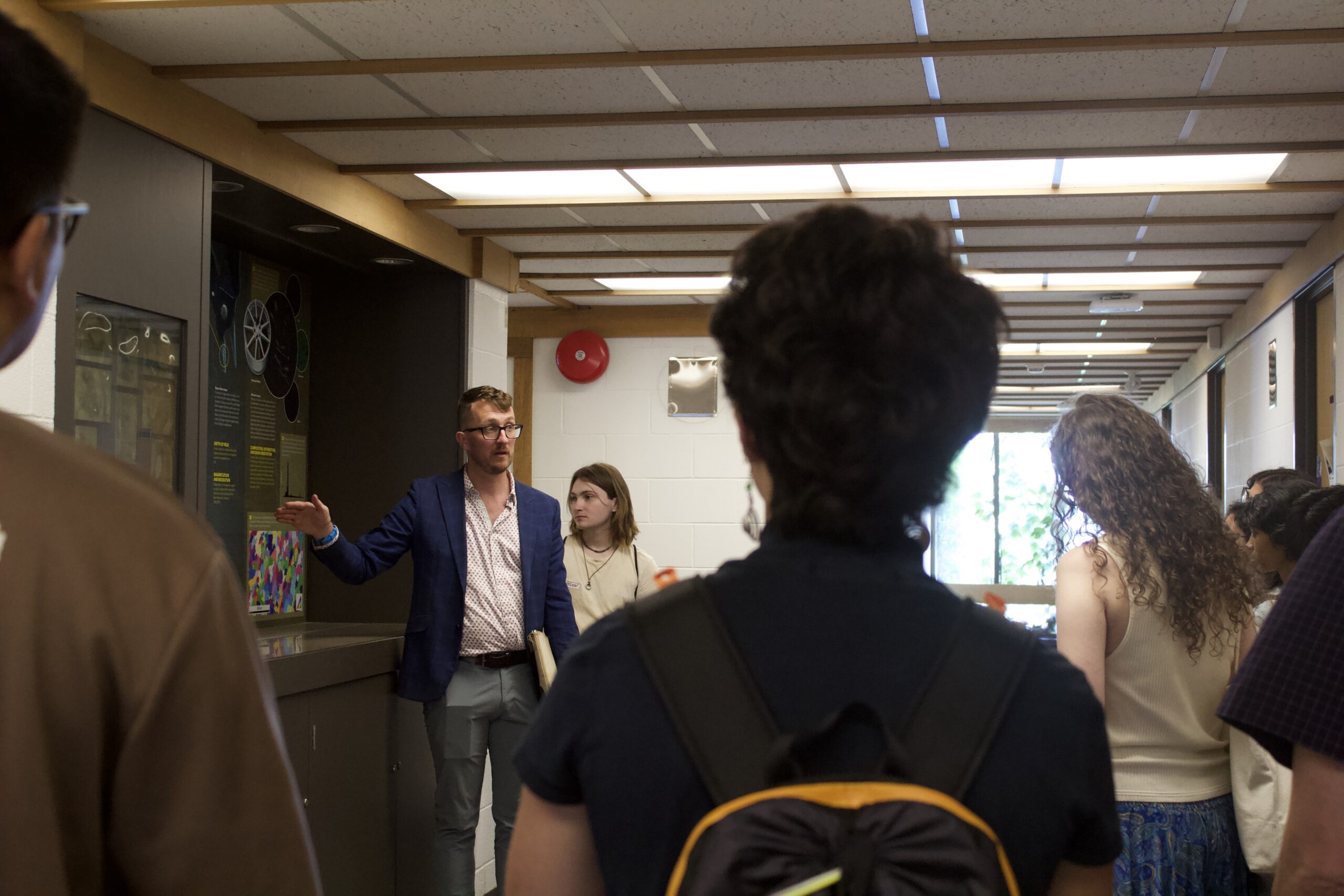
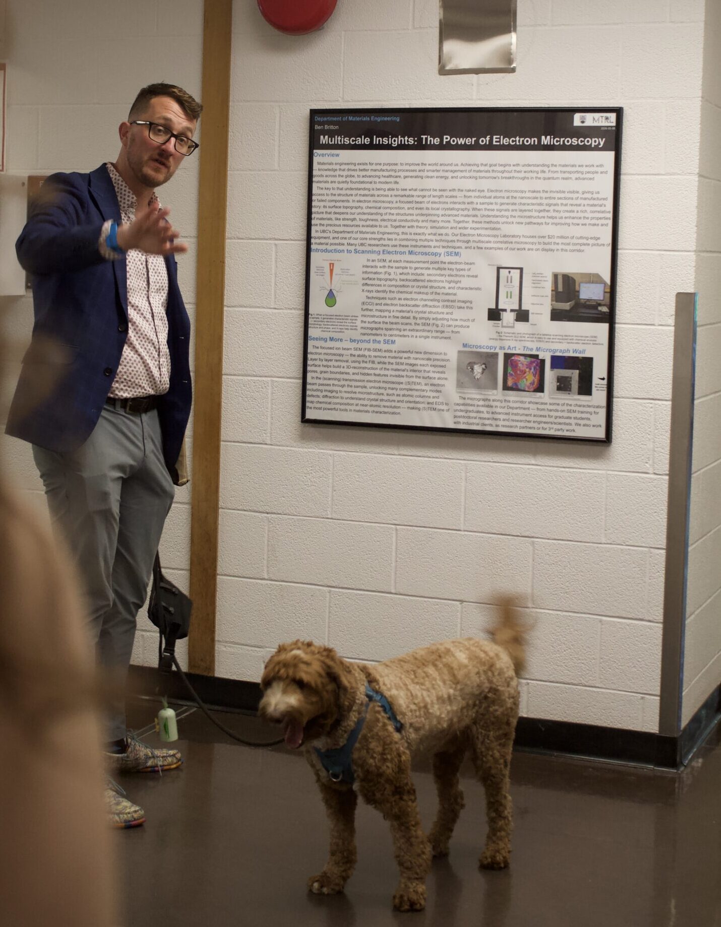
Dr. Ben Britton, a materials scientist, engineer, and Associate Professor in the Department of Materials Engineering, led the tour with his canine companion Waffles. He explained how microscopy allows researchers to observe scales beyond the limits of unaided human vision and thereby makes visible vital information about the structure and composition of different materials. This resonated with many of the research assistants, because animation is an art form that is often interested in revealing subjects and experiences that are not attainable through traditional forms of observation or recording. In a scientific context, observing materials at a microscale enables a comprehensive understanding of their characteristics, which serves the primary objective of material study– to optimize functionality and ability to predict how materials will act under different circumstances. Dr. Britton defined the concept of “material characterization,” which involves probing samples to reveal their internal structure, physical properties, and chemical composition. This is significantly different from corresponding terminology within animation, where the word “characterization” is attributed to personifying objects or materials. Similarly, in material science, the animatedness of materials is more closely linked to their properties at different scales.
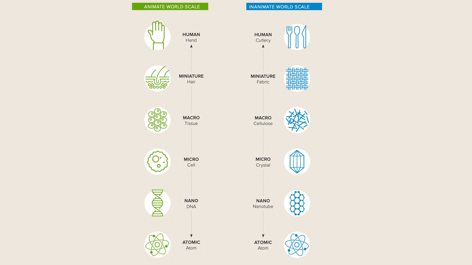
The British Royal Society’s report on animate materials, authored by Mark Miodownik, explains how materials change their qualities with scale.
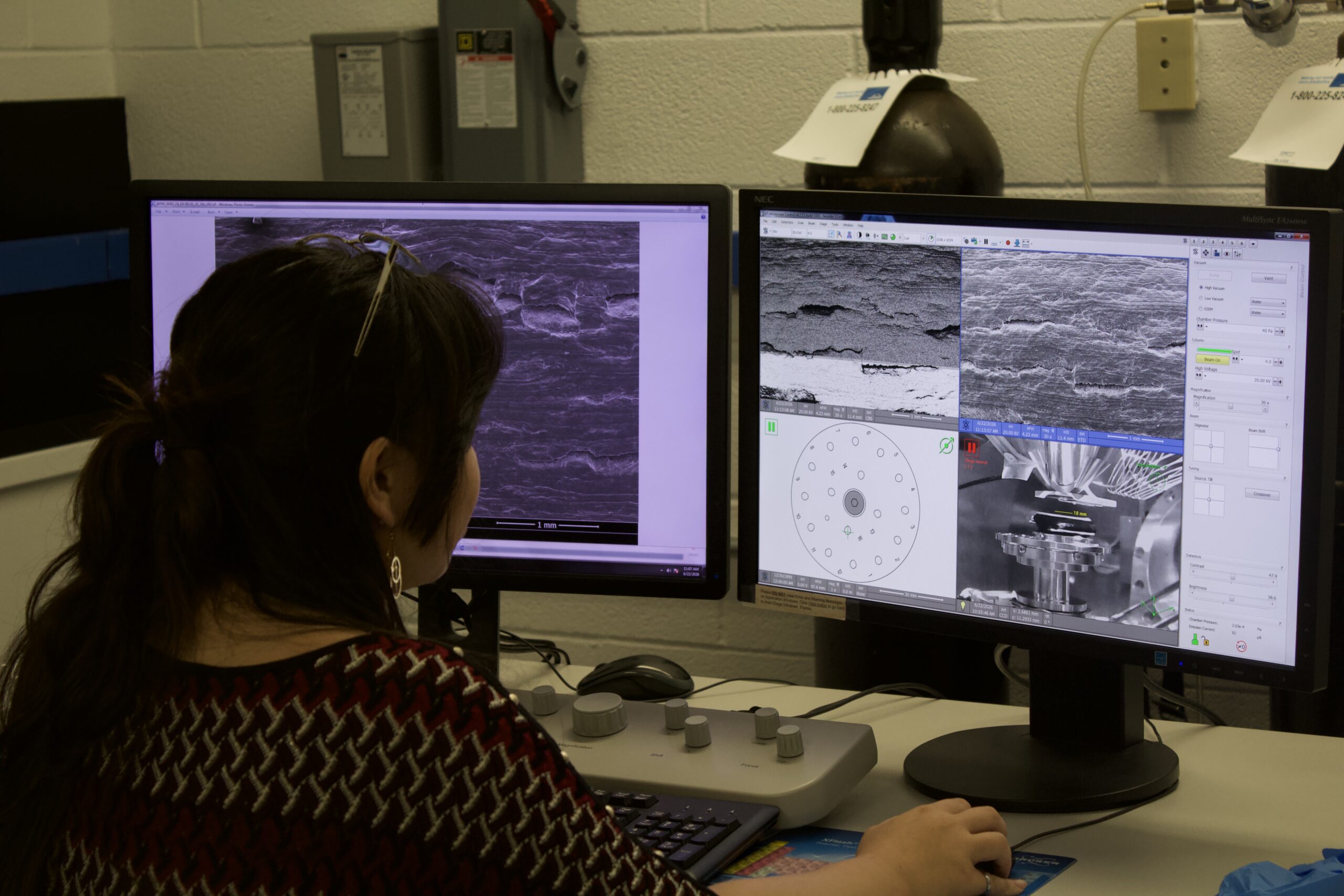
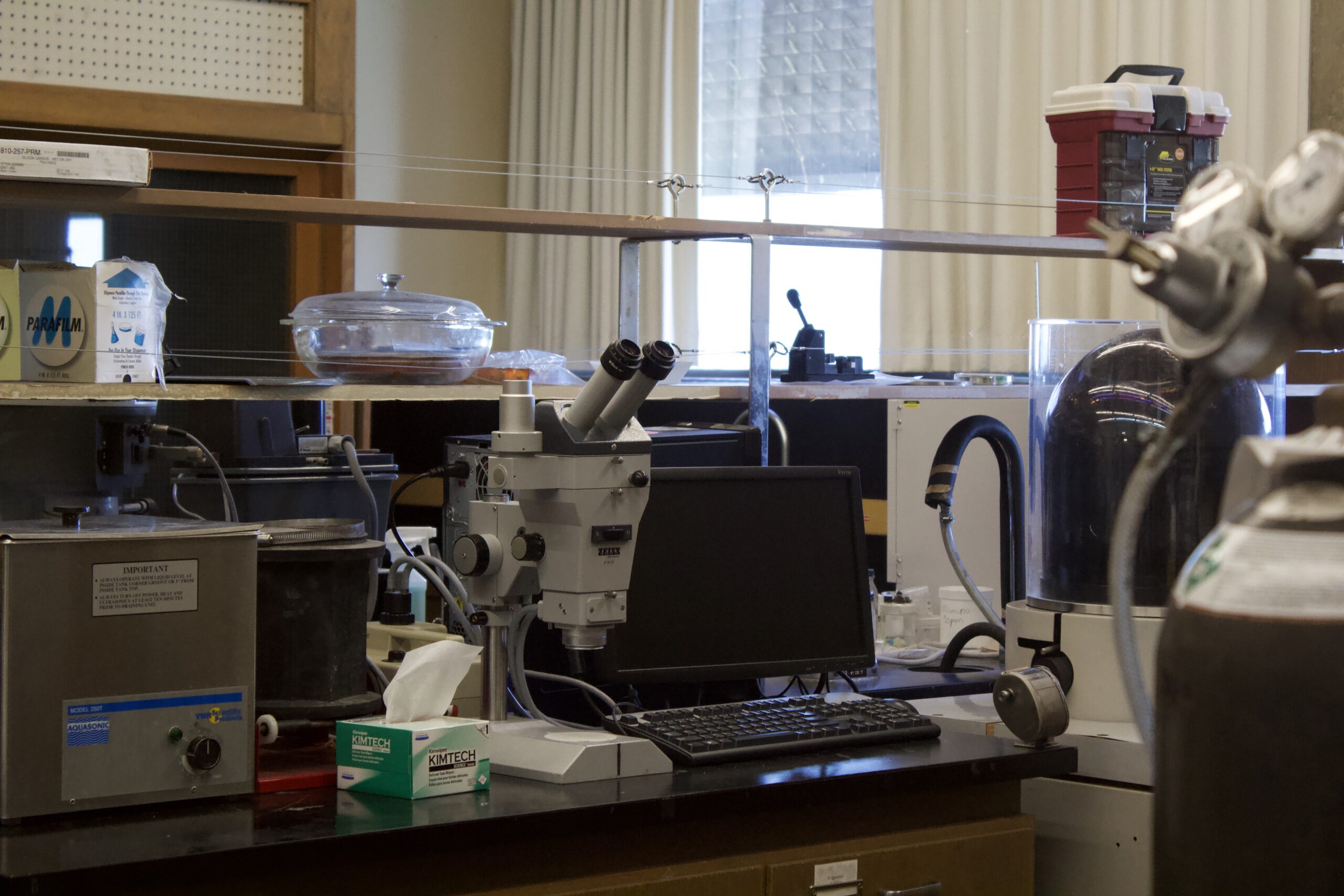
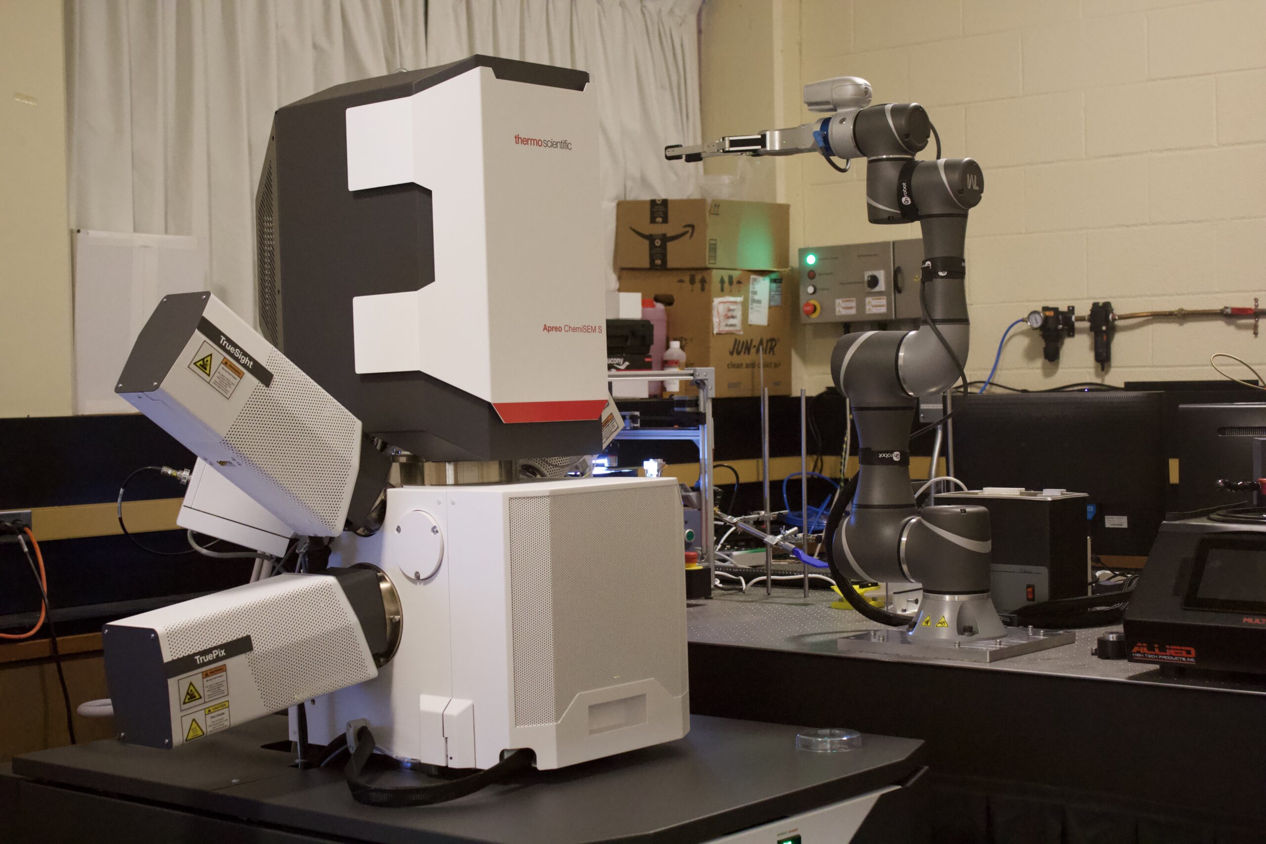
Our visit included detailed insights into the mechanics of scanning electron microscopy (SEM), in which a focused electron beam interacts with a material sample, yielding information about surface topography, chemical composition, and other properties. This knowledge facilitates improvements in characteristics such as strength, toughness, and conductivity. By adjusting the scanned area, SEM can produce micrographs ranging from nanometer to centimeter scales. Dr. Britton guided participants to the new SEM laboratory, where we observed the microscope in operation and met graduate student Jasmine Lee, who explained the process and core principles of using the equipment. As we watched a projected scan of a small aluminum sample, the nanoscale imagery reminded us of celestial landscapes. The importance of maintaining a stable environment for precise readings was emphasized, as the electron beam is highly sensitive to vibrations and stray electromagnetic fields. Even minor factors, such as acoustic vibrations or “air handling”, are carefully controlled to minimize noise in imaging data. This resonated with our experiences of preparing and carefully maintaining our animation setups, where subject stability and consistency in factors like lighting and position are crucial to the workflow; even tiny variations will be immediately visible as a sequence will flash or jitter between frames. Both scientific and hands-on animation research practices involve a bodily connection with the material and require heightened sensitivity to its controlled environment.
-
A closer view of the microscopes in operation at the SEM laboratory.
-
A closer view of the microscopes in operation at the SEM laboratory.
-
A closer view of the microscopes in operation at the SEM laboratory.
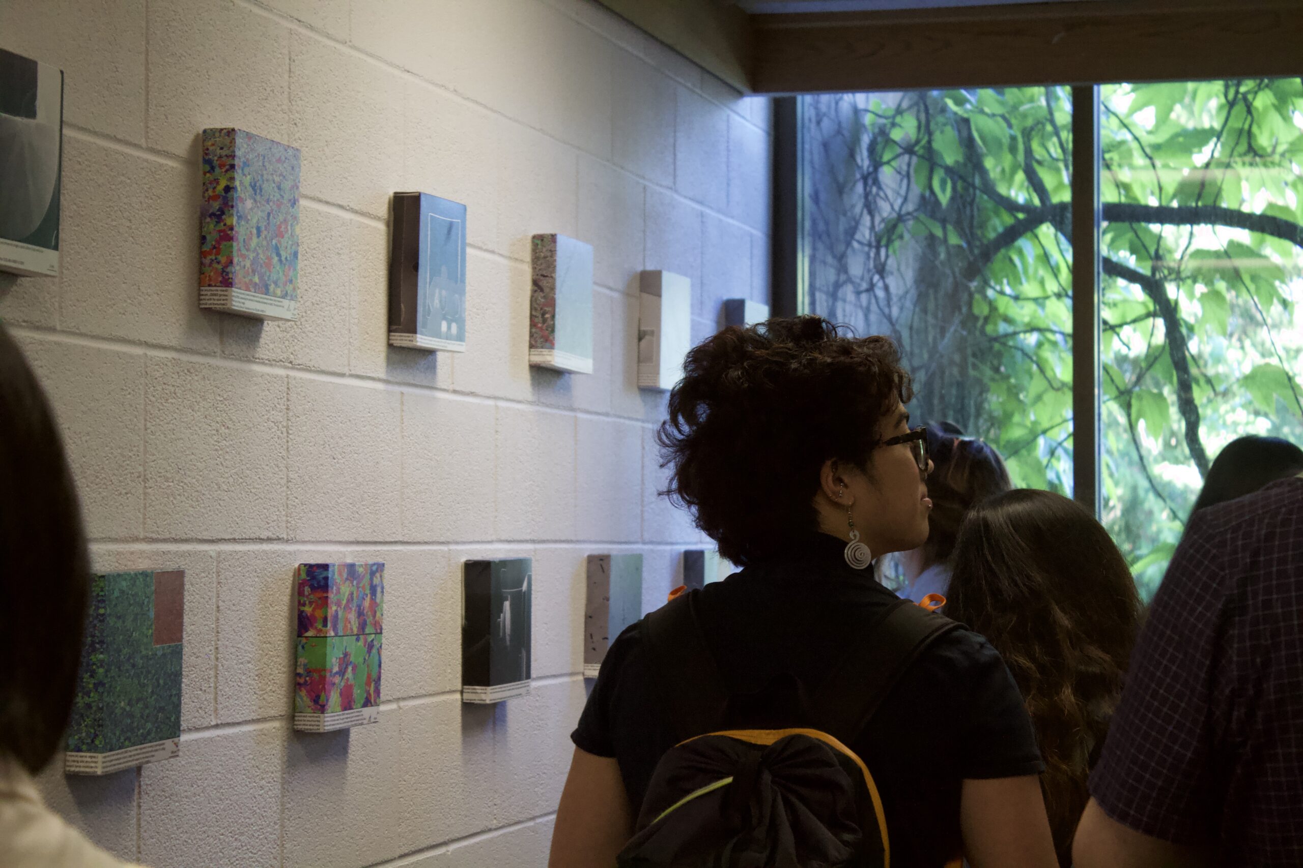
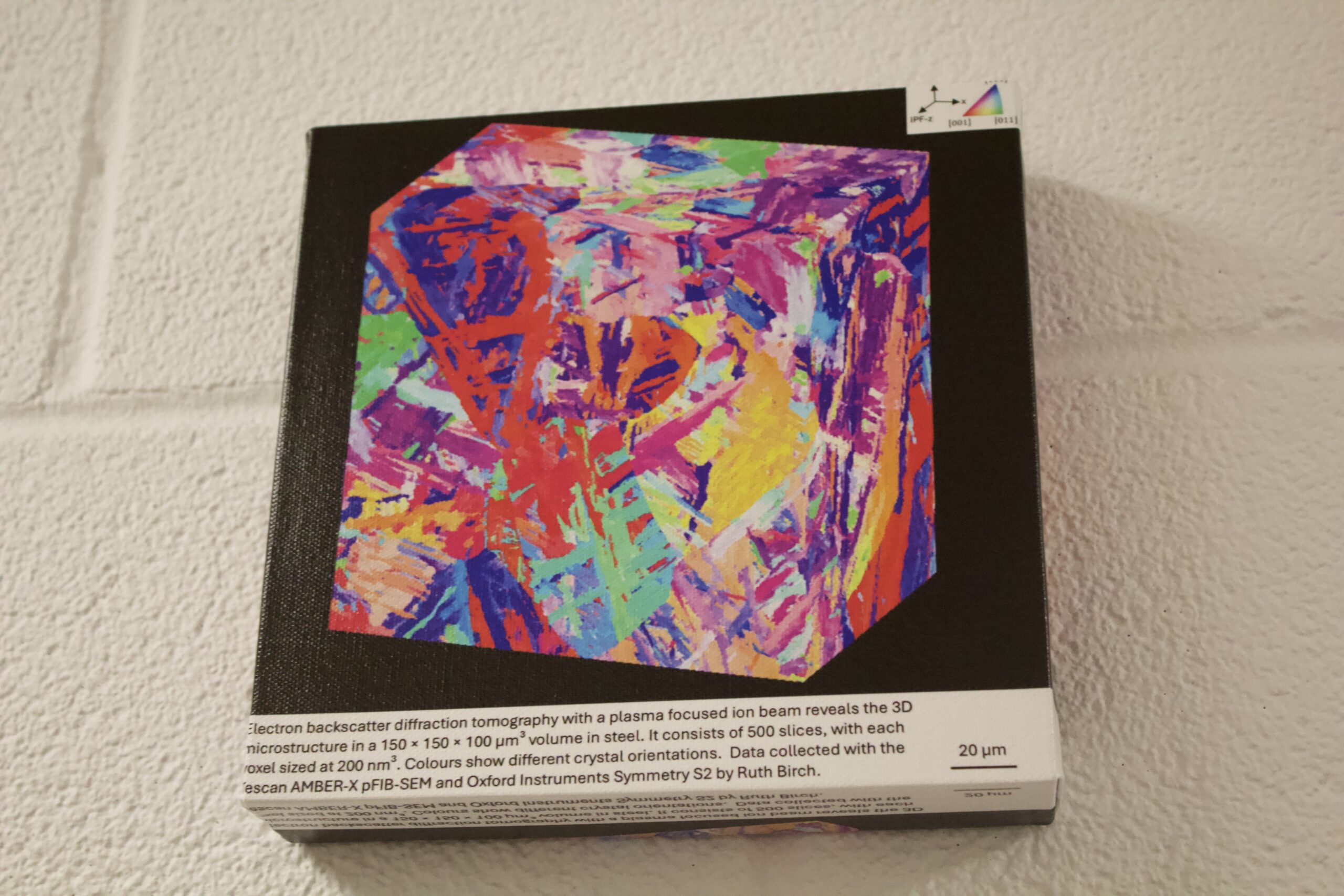
A key feature of the tour was visiting “The Micrograph Wall”, an exhibition of various micrographs generated at the laboratory that exemplified the intersection of scientific discovery and aesthetic judgement. The images resembled abstract artworks because of their visually striking colour compositions and patterns. In this discipline, colour is calibrated to enhance clarity of data rather than optical interest or pleasure; however, specific colour choices are also reliant on disciplinary aesthetic conventions. As Dr. Britton explained, “false” colour is often applied computationally to highlight specific compositional or topographical features. The underlying data remains strictly tethered to scientific observation, but the way it’s communicated visually relies on the scientist’s judgement and community standards.
-
“The Micrograph Wall” illustrates the intersection of science and art through visually striking micrographs that resemble abstract paintings.
-
“The Micrograph Wall” illustrates the intersection of science and art through visually striking micrographs that resemble abstract paintings.
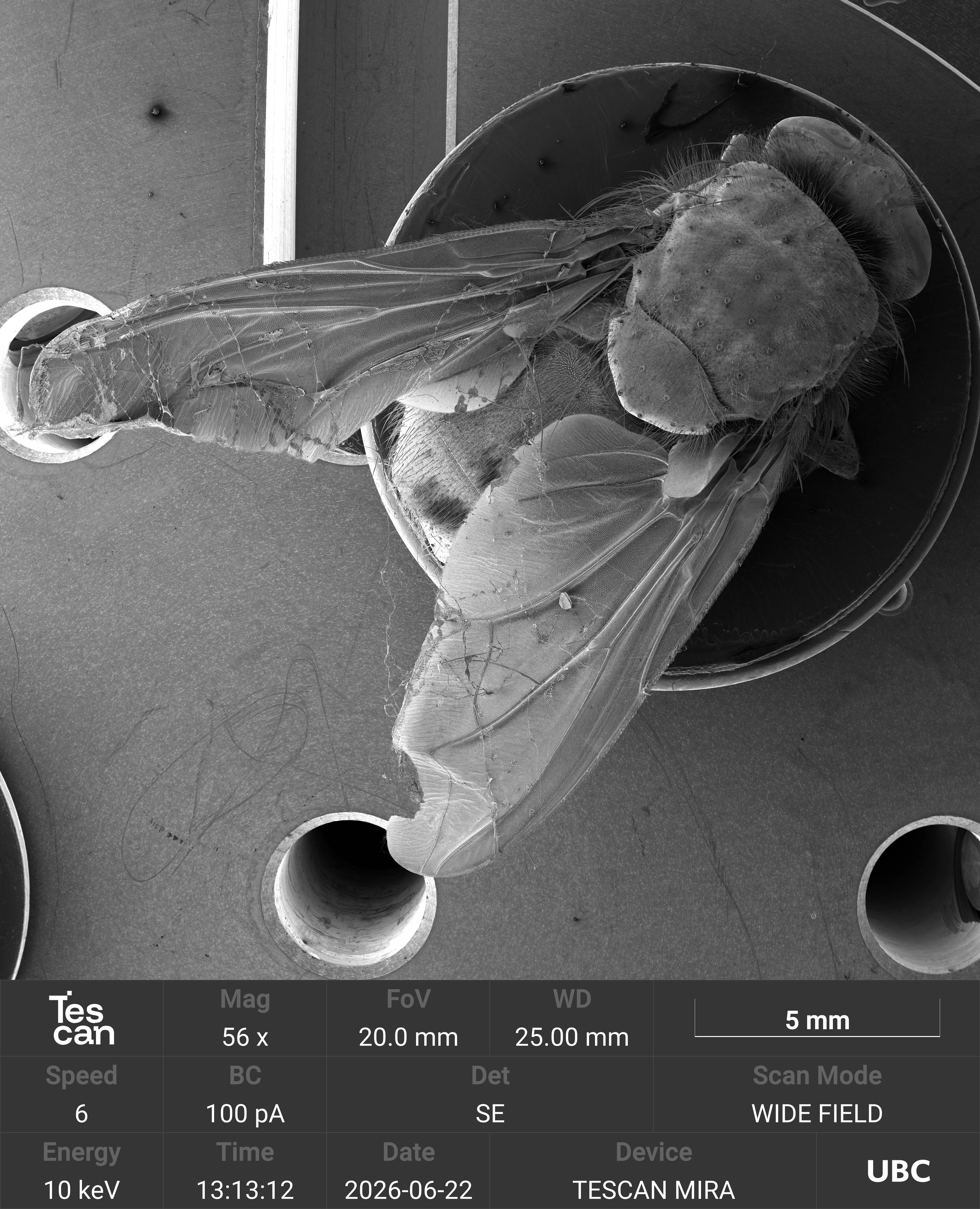
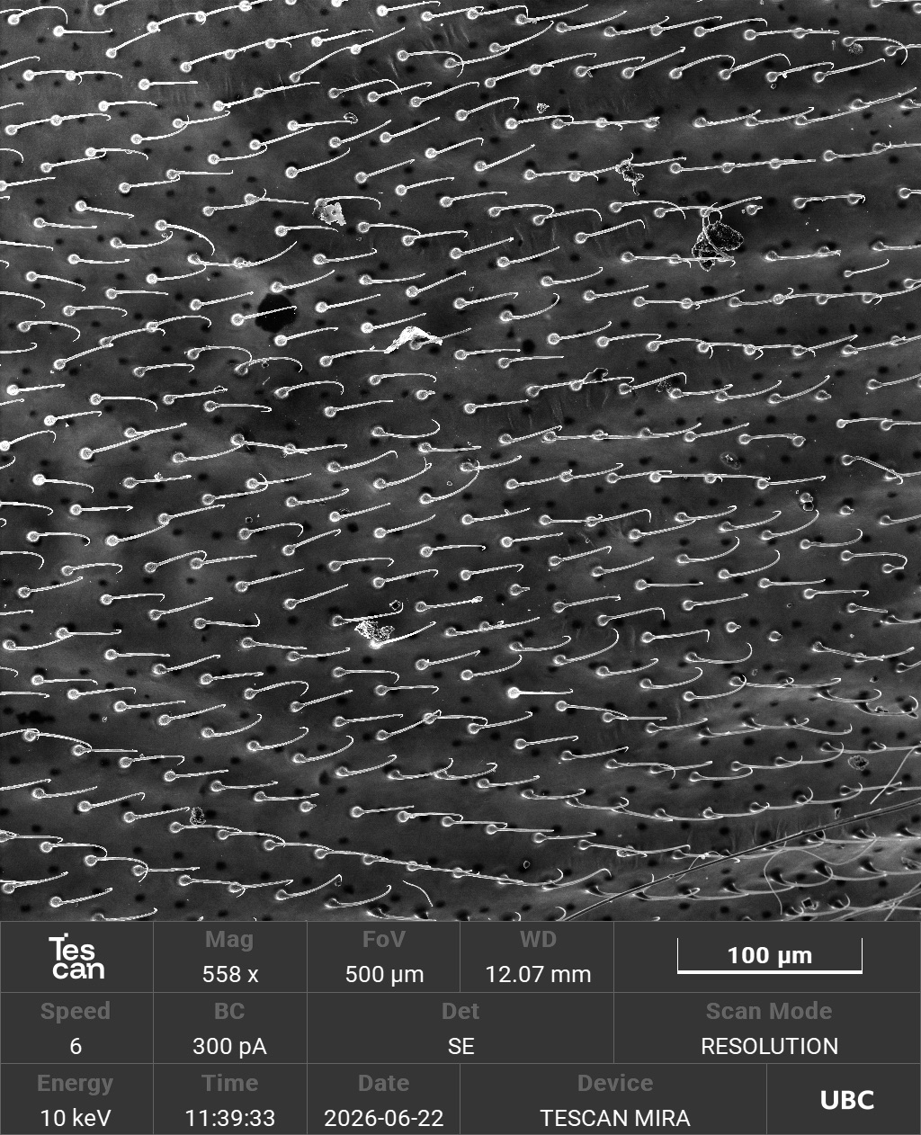
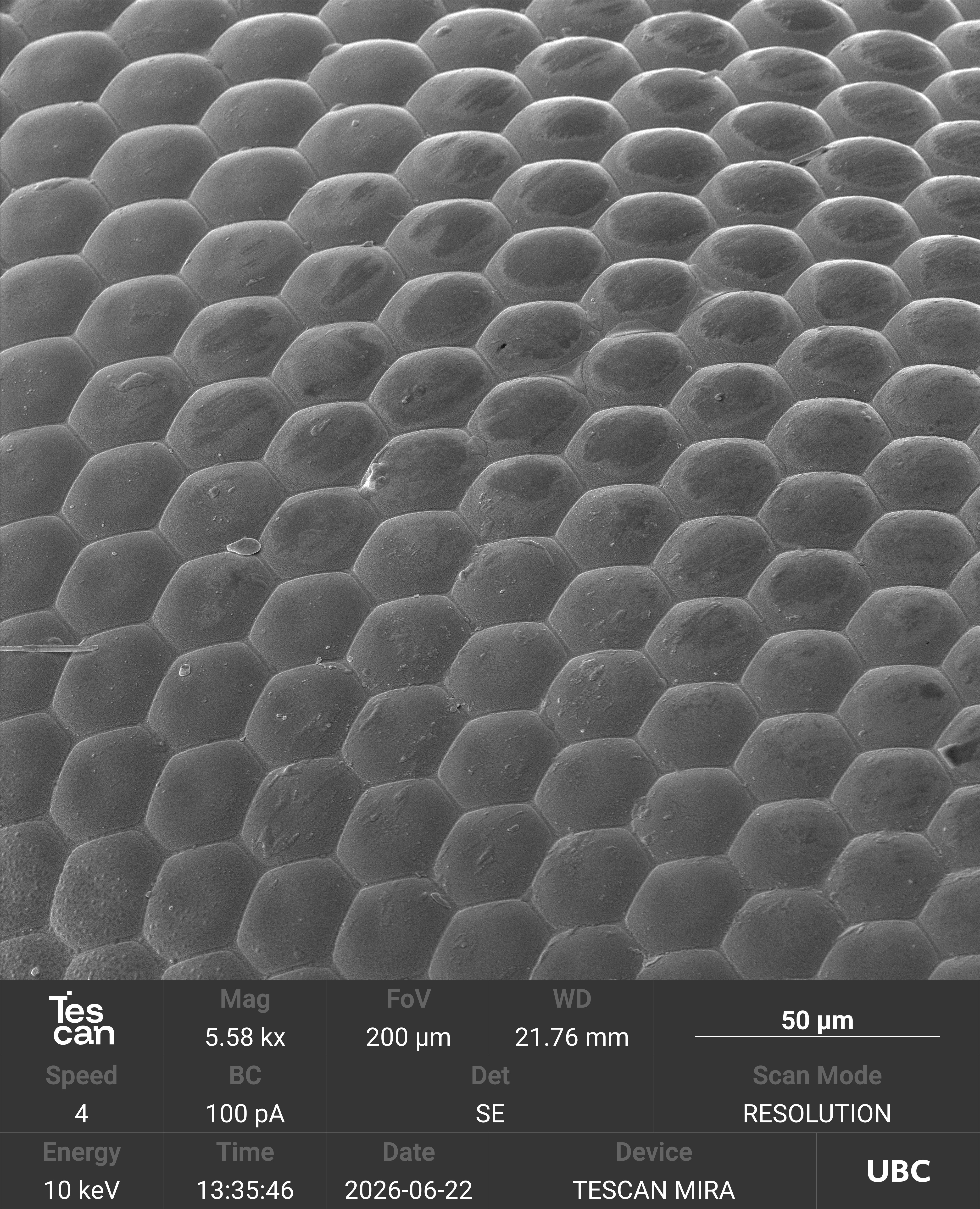
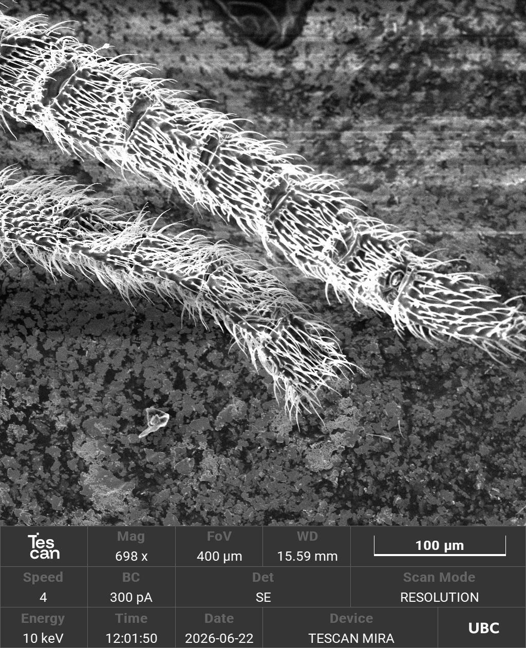
The tour concluded with a demonstration by Dr. Britton on electron microscopy, including imaging techniques, data representation, and how a focused electron beam generates data. He demonstrated live imaging of a biological specimen—a dead fly found on a windowsill of the building, coated with golden dust to render its surface appropriately conductive to electron beams. For many research assistants on the field trip, the Electron Microscope Lab was a highlight of the visit. Zen Liu likened it to weather maps that measure terrain altitudes, and noted that she’s heard of them in her high-school science curriculum as “‘an advanced microscope that you probably won’t see in your life’ kind of thing.” Zen was excited about imagining gaseous gold as a “flurry of sparkly dust.”
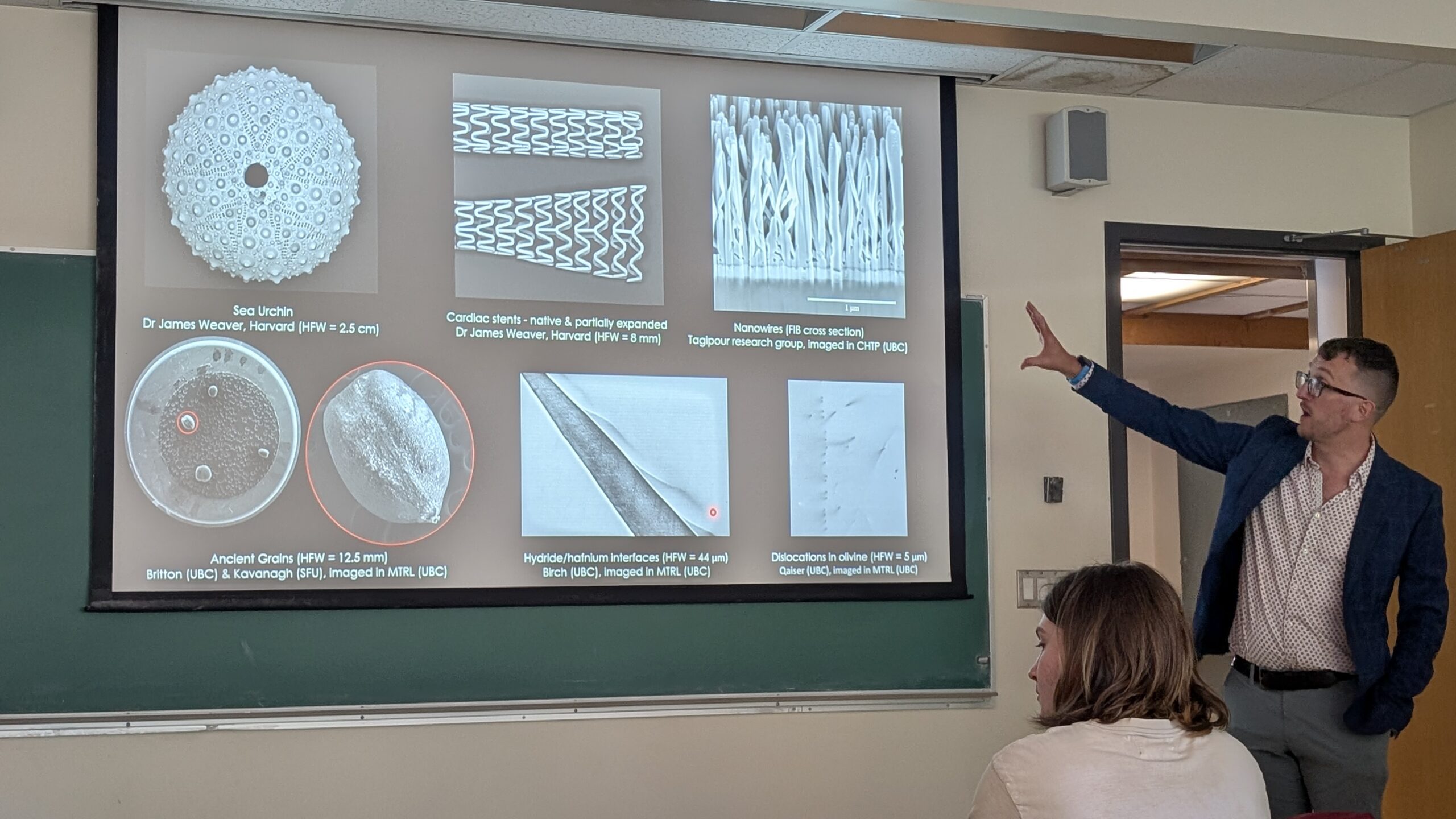
Dr. Ben Britton explains electron microscopy research, imaging techniques and data representation.
Examining materials at the microscale prompted new perspectives on molecular composition and offered a novel view of familiar objects. Participants noticed the intersection of material research through scientific and artistic lenses and saw the two merge into a more holistic approach to material research. By setting aside the technical aspects of scientific analysis that require deeper training, seeing the elements up close gave participants access to a different way of looking at material composition. Additionally, many left with exciting possibilities for gaseous gold, imagining how artists could apply it to a project and what it might look like when it interacts with other particles. Some noted the poignancy of coating a dead being with a high-value ornamental metal—echoing the dichotomy of inertness and life, and larger conversations about visibility and the subjectivity or limits of human vision that necessitate probing materials at the nanoscale. The visit ultimately showed how expertise can be communicated between disciplines.
-
Live SEM imaging of the gold-coated fly during Dr. Britton’s demonstration.
-
Live SEM imaging of the gold-coated fly during Dr. Britton’s demonstration.
-
Live SEM imaging of the gold-coated fly during Dr. Britton’s demonstration.
-
Live SEM imaging of the gold-coated fly during Dr. Britton’s demonstration.
Mengkai Zhang, a graduate student in Education at the University of Victoria with an interest in animation, joined our research field trip and similarly noted the cross-disciplinary impact of scientific visualization:
One of my greatest takeaways from the visit was gaining a clearer sense of how scientific research and humanities research (such as arts education) are increasingly dissolving the boundaries that once separated them. […] I have noticed that many concepts and methodologies traditionally associated with the arts and education are now appearing more frequently within scientific research [and communication] as well. More importantly, I had previously assumed that it was primarily the humanities that were moving toward the sciences. Yesterday, however, I suddenly realized that this movement is mutual.
MENGKAI ZHANG
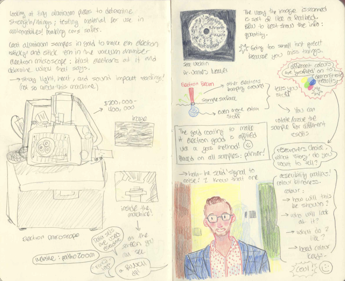
Research creation assistant Hannah Strocel’s notebook summary of the field trip.

Electron Microscopy Laboratory at UBC
Learn more about the research and training facility, including further information on their equipment on their website.
Visit the website hereScenes from the Nanoscale exhibition at MIT
Similar to The Micrograph Wall, this curated gallery of nanoscale images of materials and organisms by MIT reflects on intersections of science, nature, and artistic imagination.
See the exhibit herePathozoom
A public-access database for sharing and studying high-resolution microscopic images.
Visit the database here